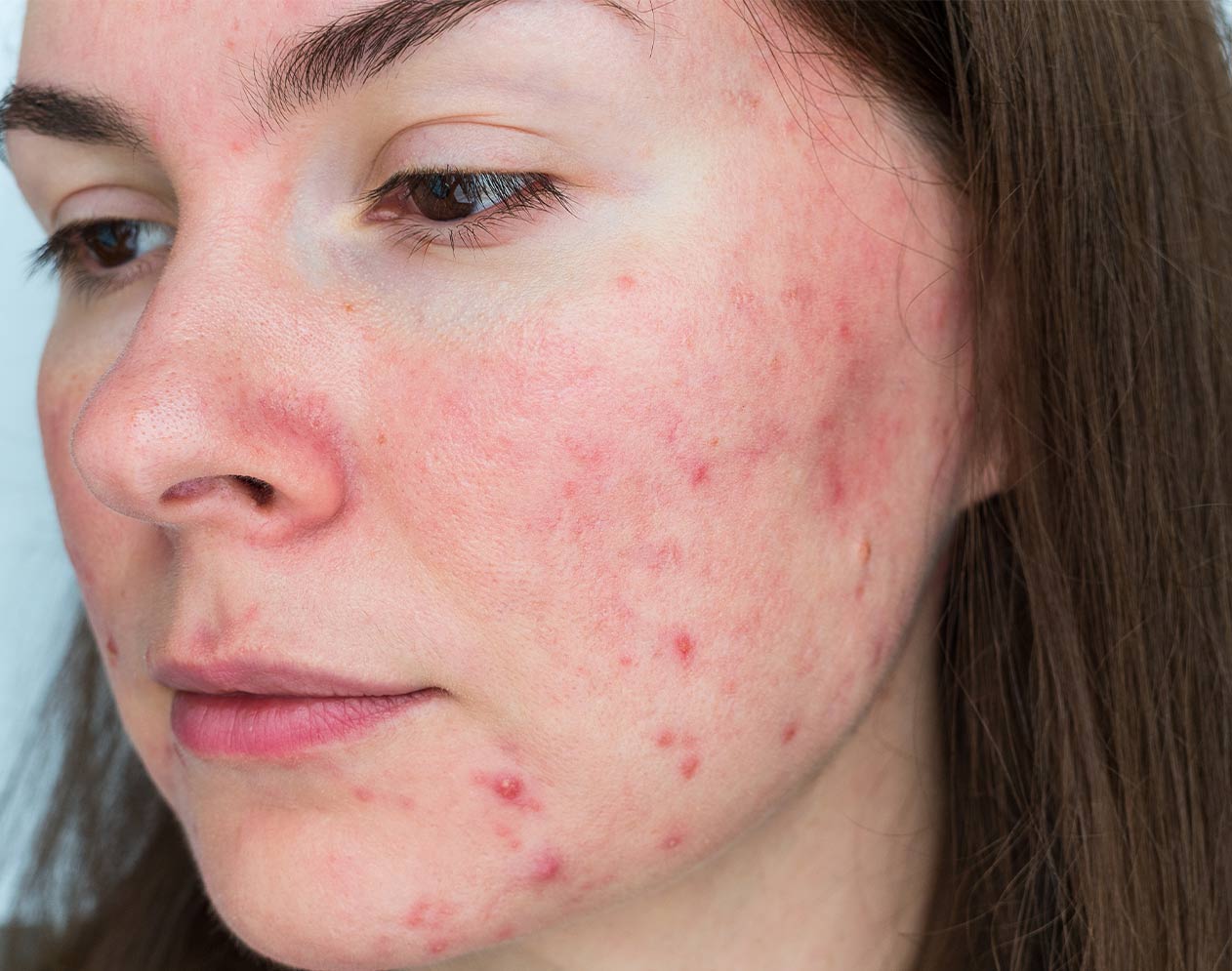
Telangiectasias are small, dilated blood vessels on the skin, often resembling a spider web.
Causes of Telangiectasias
Telangiectasias can develop anywhere on the body but are most commonly seen on the skin, mucous membranes, and sclera. Likely causes include aging, obesity, genetic factors, pregnancy, physical inactivity, and alcohol consumption. While usually harmless, they can occur as part of various conditions such as rosacea, angiomas, varicose veins, hemostasis disorders, Sturge-Weber syndrome, and many other diseases.
Treatment for Telangiectasias
The most appropriate therapeutic approach for treating telangiectasias depends on the type of lesions. Treatments include laser therapy or surgical interventions. The therapeutic approach is determined by the diameter and number of blood vessels.
For blood vessels measuring 0.1-3 mm in diameter, laser treatments with the Candela Nordlys laser, NdYAG applicator (1064 nm wavelength), or IPL PR 530 and VL555 applicators may be used, depending on clinical manifestations. The number of treatments is individualized, ranging from 3-6 sessions spaced 4 weeks apart, with visible results often after the first treatment.
Pre-Treatment Vascular Care
– Avoid tanning and tanning products for 30-60 days before treatment.
– Smokers should avoid smoking 4 hours before the treatment.
Post-Treatment Care
– Use of regenerative creams and mandatory thermal and photoprotection.
– Follow the prescribed therapy by the doctor.
Port Wine Stain
Port wine stain (Nevus flammeus) is a skin change caused by a congenital vascular anomaly, named after its color resembling bright red wine. This condition is present from birth, persists throughout life, and tends to grow proportionally with the person.
Causes of Port Wine Stain
Genetic factors play a significant role in the development of port wine stains. These changes can occur as part of syndromes such as Sturge-Weber syndrome or Klippel-Trenaunay-Weber syndrome.
Common Locations
Port wine stains are most often localized on the face but can appear anywhere on the body, particularly on the neck, upper torso, arms, and legs.
Clinical Manifestations
Lesions are typically flat and pink in appearance. As the child grows, the color may darken to deep red or purple. In adulthood, the lesion may thicken or develop small nodules.
Treatment of Port Wine Stain
Depending on the age, location of the lesion, and the extent of skin involvement, different treatments are applied.
Therapeutic options include medication, surgical therapy, and laser treatments.
At our clinic, laser treatments are performed using the Candela Nordlys laser with advanced intense pulsed light technology utilizing two applicators: PR530 and VL555. Multiple treatments are necessary, spaced 4-6 weeks apart. The effect is seen in the reduction of redness, significant regression, and even complete disappearance of the lesion.


